
The nervous system incorporates the brain and spinal cord (the central nervous system) and the network of nerves which spread throughout the body (the peripheral nervous system). Let’s quickly hash over what’s what in the nervous system.
Brain
A large organ which controls majority of the nervous system, the brain has several unique factors. One of the most important is its inability to store glucose, making it reliant on a constant flow of blood. The brain can be separated into several areas, as shown in the picture above. Each serves several different functions.
- Frontal lobe: primary motor cortex, voluntary muscle movement, some eye movements, higher thinking/reasoning, taste sensation
- Parietal lobe: primary sensory cortex (not including special senses)
- Temporal lobe: auditory and olfactory cortex, auditory association area
- Occipital lobe: visual cortex, visual association area
- Cerebellum: balance, performance on learned motor skills automatically, receives all sensations before passing them to the cerebral cortex
- Pons: bridges the cerebellum, cerebrum and brainstem together, controls respiration, sensory and motor nuclei of cranial nerves V – VIII
- Brainstem: melatonin production, part of the limbic system, controls feeding reflexes, regulate heart rate, blood pressure, respiration, digestions, body temperature, day-night cycle, some reflexes

Spinal cord
A cord made of nerve tissue, arranged into ‘tracts’ which is continuous with the brainstem, the spinal cord runs inside the spinal column, which protects it. The spinal cord links the brain to the rest of the body. It extends from the brainstem to down through the spinal column to approximately L2 in adults, and has 31 pairs of spinal nerves. These carry information to and from the spinal cord, where the posterior ascending tracts (afferent fibres) carry sensory and the anterior descending tracts carry motor impulses (efferent fibres) to muscles. The most distal part of the spinal cord is a bulbous structure called the conus medullaris. The nerve fibres which emanate from here, known as cauda equina (from Latin ‘horse tail’) innervate the pelvis, lower limbs and anal sphincter.

Meninges
The entire brain and spinal cord are protected by three membranes, known collectively as the meninges. The outermost layer, the dura mater, is a tough fibrous layer. The next layer is the arachnoid mater, which is superior to the subarachnoid space, where cerebrospinal fluid (CSF) flows. Blood vessels and nerves also travel through the subarachnoid space. Arachnoid mater gets its name from its web-like appearance. Finally, the pia mater coats the brain and spinal cord itself, and has a rich supply of blood vessels which brings nutrients to nerve tissue. The choroid plexus (which produces CSF), is also part of the pia mater, and located in the brain’s ventricles.

Cranial nerves
A set of 12 pairs of nerves which are numbered in the order of which they arise from the brain. They exit above the level of the C1 vertebrae. They are part of the peripheral nervous system and cannot be consciously controlled.
| I | Olfactory | Smell |
| II | Optic | Vision |
| III | Oculomotor | Elevate upper eyelid, pupillary constriction, most extraocular movements |
| IV | Trochlear | Downward, inward movement of the eye |
| V | Trigeminal | Chewing, clenching jaw, lateral jaw movement, corneal reflexes, face sensation |
| VI | Abducens | Lateral eye deviation |
| VII | Facial | Facial motor, taste, lacrimation, salivation |
| VIII | Acoustic | Equilibrium, hearing |
| IX | Glossopharyngeal | Swallowing, gag reflex, taste on posterior tongue |
| X | Vagus | Swallowing, gag reflex, abdominal viscera, phonation |
| XI | Spinal Accessory | Head and shoulder movement |
| XII | Hypoglossal | Tongue movement |
Of note to paramedics is;
- Damage to cranial nerve I can cause anosmia (loss of smell)
- Damage to cranial nerve II can cause sudden one sided vision loss, and may be a symptom of stroke. Sudden vision loss is always serious
- Trigeminal neuralgia and herpes zoster (shingles) commonly effect cranial nerve V, causes altered sensation or pain in the face
- Bell’s Palsy causes facial drooping and is a common stroke mimic. This occurs due to damage or infection of cranial nerve VII
- The vagus nerve (cranial nerve X) is an important cranial nerve which innervates all organs except the adrenal glands. It exerts parasympathetic control of the heart, lungs and gut – causing “rest and digest” actions. The vagus nerve is also of note as it is stimulated during Valsalva manoeuvre
Autonomic Nervous System
The autonomic nervous system (ANS) primarily controls the unconscious/automatic functions within the body, such as heart rate, digestion, respiration, pupils, urination and sexual arousal. It can be divided further into the sympathetic nervous system and parasympathetic nervous system.
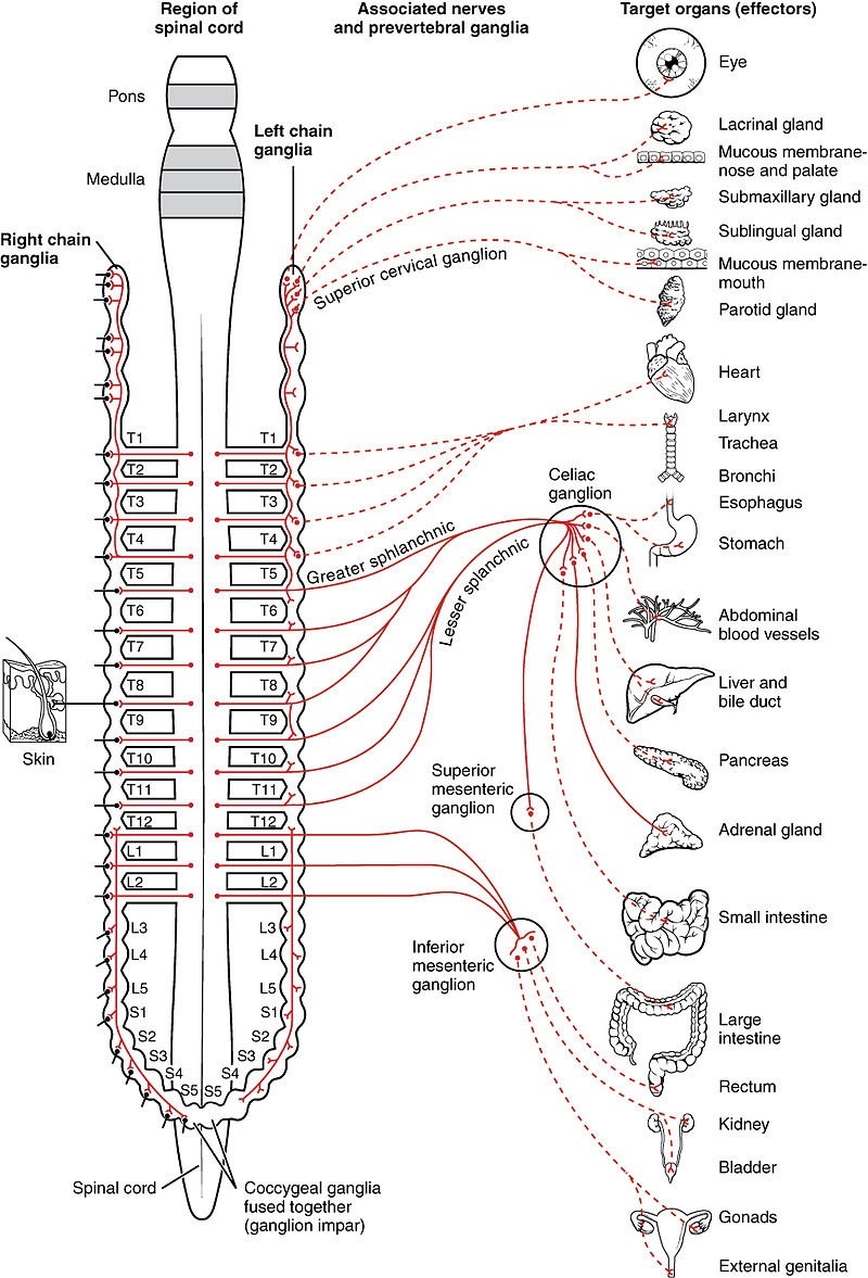
Sympathetic nervous system:
- Generally characterised as “fight or flight”, is quick to respond and generally have excitatory functions which ready the body for a threat
- Sympathetic nerve fibres originate from the thoracic and lumbar spine
- Increased sympathetic action causes pupil dilation, increased heart rate and blood pressure, increased blood flow to muscles, decreased peristalsis and blood flow to gut, and stimulates orgasm
Parasympathetic nervous system:
- Typically described as “rest and digest” or “feed and breed”, is more slowly activated and typically has a dampening effect that allows the body to rest and repair
- Parasympathetic nerve fibres originate from some cranial nerves and the sacrum
- Increased parasympathetic action causes salivation, increased blood flow to the gut, decreased heart rate and blood pressure, bronchoconstriction and sexual arousal

Cerebrospinal fluid
A specialized fluid which serves several functions, CSF is produced by the choroid plexus and circulates through the brain ventricles and the subarachnoid space. Compared to blood plasma, CSF has less dissolved protein, higher pCO2 and acidity, less glucose and more chloride ions. The functions of CSF include;
- Acts a shock absorber to protect brain and spine
- Supports the brain by allowing it to float, reducing the weight
- Transports nutrients, chemical messengers and waste products
- Maintenance of intracranial pressure (ICP), as production and reabsorption of CSF can compensate for small changes
Bottom Line
The brain represents the main control centre for all other body functions. It connects to the vital organs and muscles through cranial and spinal nerves – any disruption or damage to this system can cause critical illness and death.