Head trauma is a significant contributor to trauma deaths worldwide and is the leading cause of disability in people under 40. Although we usually talk about traumatic brain injuries (TBIs), given the lack of a CT scanner on most ambulances, we’re going to talk about traumatic head injury in this article.
Epidemiology
Causes:
- Falls (42%)
- MVAs (29%)
- Assault / non-accidental injury (14%)
- Sporting clashes (most commonly footy)
Outcomes:
- Concussion / brief LOC (60%)
- Ongoing disability (35%)
- Death (4.6% of all hospitalised TBI patients)
Patient Presentation
Can be incredibly varied, depending on severity of mechanism and patients baseline health status
Generally:
- Confusion
- Amnesia and repetitive questioning
- Headache
- Nausea
- Irritability
- Drowsiness
More concerning symptoms:
- Pupil abnormality
- Multiple vomits
- Seizure
- Unconscious episode > 5 minutes long
- Obvious skull fracture/deformity
- Neurological deficits
- Bradycardia, hypertension and irregular respirations (Cushing’s Triad)
Pathophysiology
- External force acting on the head causes injury to the underlying brain tissue
- Can be broadly categorised as closed head injury (blunt), penetrating head injury or blast injury
- Blunt injury causes the brain to jolt around in the head (coup contrecoup injury), causing compression of the soft brain tissues. Remember that the brain is floating inside the skull in a pool of cerebral spinal fluid, so its fairly mobile. Direct damage to neurons and brain vasculature occurs, but symptoms can be quite variable depending on which areas of the brain are affected. Some injuries will result in focal contusions, whilst others will cause diffuse (widespread) damage. Its worth noting that the injury is not isolated to the site of the impact. In what’s known as a coup-contrecoup injury, the skull will rattle around in the skull causing damage to the opposite side of the impact.

- Penetrating injuries, as the name suggests, occurs when a foreign body penetrates the skull and directly damages brain tissue and blood vessels. In addition to brain injury, major haemorrhage is an immediately life threatening concern. Subsequent infection is also common
- Blast injury occurs when the extremely high pressure shock wave from a blast reverberates inside the skull, causing wide spread disruption to brain tissue and blood vessels. Its often accompanied with other blast injuries to hollow organs, eyes and ears
- Each of these three categories result in the primary head injury – the damage that occurs at the time of the trauma. Unfortunately nothing can be done to modify the primary head injury
- Secondary head injury occurs subsequent to the initial insult and is affected by many factors. Preventing or reducing secondary head injury is the main goal of paramedics
- Multiple underlying mechanisms impact secondary head injury. Some of these include changes to nerve depolarisation, mitochondrial dysfunction, release of inflammatory mediators, axonal degeneration and cell death. Whilst these are areas of research, currently we have no way of modifying these
- Factors which we can impact are: cerebral perfusion pressure, oxygenation, ventilation and blood glucose. Hence our out-of-hospital management revolves around optimising these features. These will be addressed individually in the Management section.
- After the initial insult, inflammation and tissue oedema occur in the brain. There may also be bleeding present. As the skull is a fixed volume, these factors lead to increased intracranial pressure (ICP)
- This raised ICP leads to compression of the brain’s blood vessels and reduced cerebral blood flow. Homeostatic mechanisms kick in, though these have limited ability to compensate (but more on that in a minute). Decreased cerebral blood flow results in hypoxia and death of brain tissue, which causes further swelling. This worsening cycle of cell death, oedema and raised ICP results in compression of the brain stem and respiratory centre
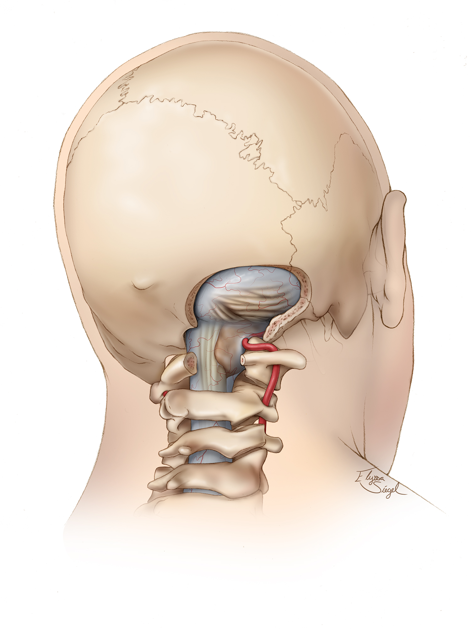
- This occurs as the only way out of the skull is the foramen magnum, which is where the spinal cord connects to the brainstem. As pressure builds, the brain tissue is forced downward through the foramen magnum, a process known as “coning”

- The disruption of the brain stem leads to Cushing’s Triad (hypertension, bradycardia, irregular respirations)

- By this stage, damage is catastrophic. Respiratory dysfunction causes CO2 retention, which results in vasodilation and further increased ICP. This will lead to further respiratory depression, bradycardia and cardiac arrest.
Hang on – our body loves homeostasis… why doesn’t it do something about raised ICP?
Our body does love homeostasis! So we’re not totally helpless. Remember our neuro anatomy – the brain is floating inside the skull in a nice pool of cerebrospinal fluid (CSF). In the event of raised ICP, our very clever body will reabsorb CSF to make more room for swelling or bleeding. However this is quite limited, as CSF only makes up 10% of cranial volume. Once all the CSF has been reabsorbed, if the source of raised ICP has not been resolved, the patient will continue to deteriorate.
This homeostatic mechanism can be viewed in stages, as described by the Monroe-Kellie Hypothesis.

You mention that we can control cerebral perfusion pressure… how do I do that?
Our brain is very energy hungry, and is sensitive to changes in perfusion. We describe the flow of nutrient rich blood to the brain as cerebral perfusion pressure (CPP). CPP is calculated at the mean arterial blood pressure (MAP) minus the intracranial pressure (ICP).
CPP = MAP – ICP
Basically for the brain to get nutrients, your average blood pressure needs to be higher than the pressure inside the brain, forcing nutrients into it. In a healthy brain, ICP is roughly 5-15mmHg, and we know that a healthy level of cerebral perfusion is > 60mmHg, therefore we typically want the patient’s MAP to be > 70mmHg.
Just to prove it to you
75mmHg (MAP) – 10mmHg (ICP) = 65mmHg (CCP)
That’s a happy brain!
Now I mentioned earlier on, insult to the brain and the subsequent swelling/bleeding causes an elevated ICP. Therefore if we want to keep the brain properly perfused, we’ll have to increase our patient’s MAP. We will do this by providing IV fluid resuscitation (or blood products if available) to raise SBP > 120mmHg. This will result in higher MAP, which will help overcome the elevated ICP.
However we have another trick up our sleeve! Remember back to your anatomy and physiology training – high CO2 levels cause vasodilation, whilst lower CO2 levels cause vasoconstriction. Therefore by keeping the patient’s CO2 levels at the lower end of normal range we can help control blood vessel diameter and manage CPP. Don’t worry – there’s more on this in the Management section!
A final tip for managing ICP is position – a very low level of evidence suggests that a 30o head tilt will improve venous drainage and reduce ICP. However this may not be practical for all patients and may impact spinal immobilisation, so its not included in Victorian CPGs.
Assessment:
- Primary Survey
- Trauma secondary survey
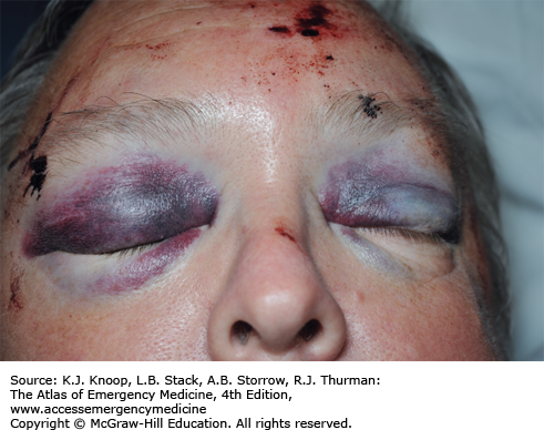
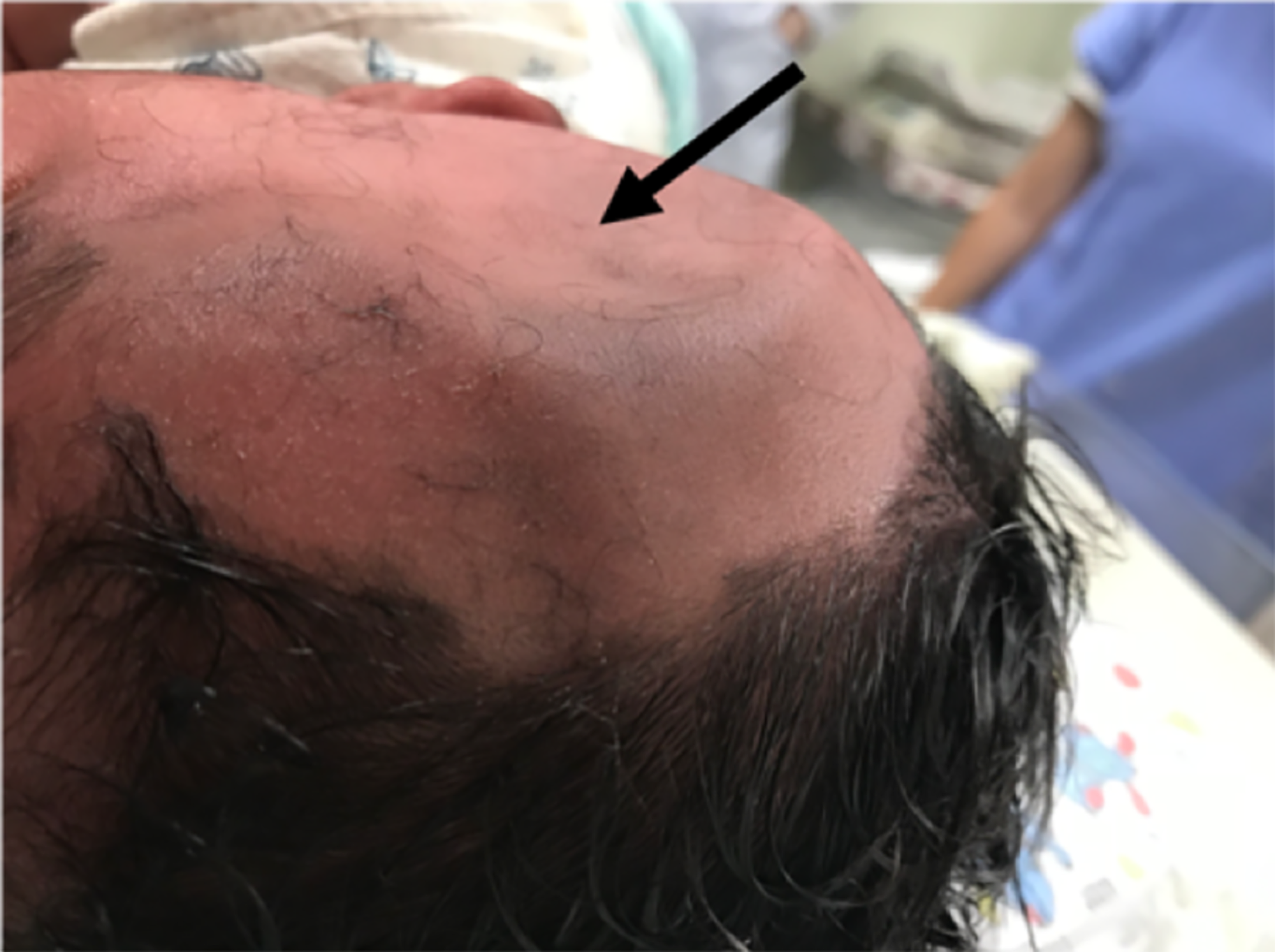
- Assess for skull fracture – boggy mass, depressed skull, Battle Sign, periorbital ecchymosis (“raccoon eyes”) or CSF leaking from ears/nose
- Consider potential for C-spine injuries, as any significant injury to head likely also impacted C-Spine
- Once major head injury is identified, don’t forget to thoroughly check for other injuries to the body


- VSS
- Including BGL for all altered conscious state patients
- AMPLE history
- Including considerations for alcohol and other drug use
- Good understanding of mechanism is important to risk stratification
- Relevant medical history including previous neurosurgery or head injuries
Assessment Pearls
- Elderly patients may sustain significant head injuries from seemingly simple falls from standing height
- Patients on anti-coagulants (Rivaroxaban, Warfarin etc) are at increased risk of serious intracranial bleeding. Those on anti-platelet agents (Aspirin, Clopidegrol) are also at increased risk, although not as much as anti-coagulants
- In head injured, intoxicated patients – always assume low GCS is the result of head injury, rather than intoxication
Differential Diagnoses
- Skull fracture
- Subdural haematoma
- Epidural haematoma
- Subarachnoid haematoma
- Cerebral contusion
- Diffuse axonal injury
Time Criticality
Use Trauma Time Critical guidelines to help guide decision making
- Actual Time Critical (deranged vital signs)
- HR < 60 or > 120
- RR < 10 or > 30
- SBP < 90
- SpO2 < 90%
- GCS < 13
- Emergent Time Critical (pattern of injury)
- Penetrating head trauma
- Significant blunt trauma to two or more body areas
- Serious blunt trauma to a single body region (use 5HEDS mnemonic)
- > 5 minutes LOC
- Head (skull) fracture
- Emesis ≥ 2 times
- (Neurological) deficit – this may be a result of serious head injury or suspected spinal cord injury, which is also time critical
- Seizure of any duration
- Potential Time Critical (mechanism of injury)
- Motorcycle or cyclist impact > 30km/h
- Motor vehicle accident > 60km/h
- Pedestrian struck by vehicle
- Ejection from vehicle
- Prolonged extrication (typically > 20 min)
- Fall from height > 3m
- Struck on head by object falling > 3m
- Explosion
AND
- Age < 12 or > 55, OR
- Pregnant, OR
- Significant underlying medical condition
Paediatric Time Criticality
Melbourne’s Royal Children’s Hospital primarily uses GCS to risk stratify paediatric head injuries, as shown here

Where risk factors are:
| o Severe headache o Persistent altered mental status/acting abnormally o Abnormal neurology o Suspected child abuse o Palpable skull fracture | o Signs of base of skull fracture o Non-frontal scalp haematoma (occipital, parietal or temporal) in child <2 years o Severe mechanism o Post-traumatic seizure o Loss of consciousness o Persistent vomiting | o Known bleeding disorder/anticoagulation o Ventriculoperitoneal shunt o Neurodevelopmental disability |
Actual Time Critical (deranged vital signs)

Emergent Time Critical (pattern of injury)
- Penetrating head trauma
- Significant blunt trauma to two or more body areas
- Serious blunt trauma to a single body region
- Signs of skull fracture
- Neurological deficit – this may be a result of serious head injury or suspected spinal cord injury, which is also time critical
- Seizure without full recovery
NOTE: There is no reference to 5HEDS for paediatric patients, so this is drawn from RCH guidelines
Potential Time Critical (mechanism of injury)
| Ambulance Victoria CPG | Royal Children’s Hospital CPG |
|---|---|
| o Motorcycle or cyclist impact > 30km/h o Motor vehicle accident > 60km/h o Pedestrian struck by vehicle o Ejection from vehicle o Prolonged extrication (typically > 20 min) o Fall from height > 3m o Struck on head by object falling > 3m o Explosion | o MVA with patient ejection or rollover, death of another passenger o Pedestrian impact or cyclist struck by vehicle o Fall ≥ 1m (age < 2 years) o Fall > 1.5m (age ≥ 2 years) Head struck by high impact object |
Paramedic Management of Traumatic Head Injury
Care Objectives:
- Identify and appropriately triage potentially serious head injury
- Prevent secondary brain injury by optimising ABCs
I recommend using an ABCDE approach to these patients. Note these should be prioritised in order of need and where possible implemented concurrently
A – AIRWAY
- If the patient’s airway is patent, don’t mess with it. Causing the patient to gag will result in raised ICP and potentially worsening head injury
- If airway is not patent, consider
- Position laterally
- Triple airway manoeuvre
- Suctioning as required
- If airway still not patent, insert nasopharyngeal airway (NPA)
- If unable to insert NPA / airway still not patent or GCS < 10, MICA to perform rapid sequence intubation
- If MICA not available / RSI not possible and gag reflex is absent, insert supraglottic airway (SGA)
B – BREATHING
- Optimise ventilation and oxygenation, aiming for targets
- Vt 6 – 7mL/kg
- SpO2 > 95%
- EtCO2 30 – 35mmHg
- Address causes of hypoxia and avoid hypo/hypercapnia
- Consider chest decompression in multi-trauma
PRACTICE POINT: hypercapnia causes vasodilation, which would lead to increased cerebral blood flow (CBF) and contribute to raised intracranial pressure (ICP). In the paramedic setting we cannot accurately measure ICP, but we assume it is normal or elevated in patients with head injuries. Therefore we target a lower than normal range for ETCO2. However it is also worth noting that hypocapnia (leading to vasoconstriction) can also contribute to secondary brain injury. Also ETCO2 (which we measure) is not quite equivalent to the arterial partial pressure of CO2 (PaCO2) in the blood – it is usually 3.8mmHg lower than PaCO2. However in TBI this gradient is even greater, and its further complicated if chest injuries are also involved. It is for these reasons we aim for a ‘low normal’ ETCO2 of 30 – 35mmHg.
C – CIRCULATION
- Find and control external haemorrhages using haemostatic dressings and tourniquets as necessary
- Apply pelvic binder if blunt trauma to pelvis or unconscious/hypotensive trauma patient
- Aim for SBP > 120mmHg in patients with head injury
- Fluid resuscitate with Normal Saline 250mL boluses to a maximum of 40mL/kg
- Consider access to blood products and use them where available (contact DM to facilitate)
- MICA: if there is inadequate response to fluid resuscitation, and other causes of hypotension have been excluded, consider adding Metaraminol boluses or Noradrenaline infusion
- MICA: Give Calcium Gluconate 4.4mmol (2g) over 2-5 min after 4 units PRBC or if iCa < 1.12mmol/L at any stage. If iCa < 1.12mmol/L after initial dose, give further Calcium Gluconate 2.2 – 4.4mmol (1-2g) IV over 2-5 min.
Let’s talk about that blood pressure goal! Earlier on we discussed cerebral perfusion pressure (CPP) and what we’re trying to achieve. Healthy CPP is > 60mmHg, and CPP is calculated as mean arterial pressure (MAP) minus ICP.
So, how does this systolic blood pressure fit in? Lets compare two examples…
Example 1: Head injured patient without fluid resuscitation
ICP = 25
BP = 105/65 (MAP = 72)
CPP = 72 – 25
CPP = 47 Poor cerebral perfusion
Example 2: Same head injured patient, fluid resuscitated to a goal of SBP > 120
ICP = 25
BP = 132/70 (MAP = 91)
CPP = 91 – 25
CPP = 66 Acceptable cerebral perfusion
D – DISABILITY
- Manage seizures with Midazolam as per CPG A0703 (Seizures)
- Treat pain with analgesia as per CPG A0501 (Pain Relief)
PRACTICE POINT: Which analgesia is best? Lets dissect the options.
Morphine = effective opioid analgesia, however has a tendancy to cause nausea, sedation and hypotension. None of which is ideal in head injury.
Fentanyl = effective opioid analgesia, with less side-effect profile than Morphine. I tend to use this.
Ketamine = effective dissociative anaesthetic agent, however can cause hypertension and emergence phenomena. I would consider consultation if Fentanyl was not sufficient.
Paracetamol = mild, non-opioid analgesia. Probably suitable for fully conscious head injuries, but shouldn’t be given to patients in an altered conscious state.
Methoxyflurane = effective inhalational anaesthetic agent, but wears off quickly and requires the patient to be fully conscious. Not suitable for unconscious head injuries.
- Manage mild agitation with judicious opioid analgesia. Avoid using Midazolam for agitation due to its impact on conscious state and airway reflexes
- If agitation is so severe that it prevents treatment, treat using Ketamine IM as per CPG A0708 (Acute Behavioural Disturbance)
- Consider an anti-emetic prophylactically, as vomiting will cause ICP to spike
- Manage hypoglycaemia with IV 10% Dextrose or IM Glucagon as per CPG A0702 (Hypoglycaemia)
E – ENVIRONMENT / ESCALATION / EVERYTHING ELSE
- Maintain normothermia
- Trauma blanket bundle
- Vehicle heater on
- Minimise exposure
- Note the goal is normothermia – T > 39oC has been found detrimental in head injury
- Consider spinal immobilisation and apply C-collar where indicated
- Dress wounds with appropriate dressings
- Escalate trauma time critical head injuries to MICA
- Consider HEMS if > 60 min from major trauma service
- Re-assess throughout transport
- Head injury observations 15/60 for 2hrs
Hospital Destination
Time Critical Head Injury = Major Trauma Centre within 60 minutes travel time
Minor – moderate head injury = nearest ED with imaging support
Bottom Line
Head injuries can be devastating. GCS on arrival is a key indicator of severity, but its not the only one. Assess for 5HEDS and mechanism of injury in all patients to avoid missing those who are time critical. Optimise ABCDE to reduce the risk of secondary brain injury. Transport without delay to an appropriate facility.
References
https://www.aihw.gov.au/reports/injury/hospital-separations-brain-injury-2004-05/summary
https://aneskey.com/wp-content/uploads/2016/06/m_atl_ch1_f003.png
https://upload.wikimedia.org/wikipedia/commons/thumb/0/09/Contrecoup.svg/1200px-Contrecoup.svg.png
https://assets.cureus.com/uploads/figure/file/66974/1d2936a070e611e9a8e733857e32f951-FIGURE-1.png
https://www.osmosis.org/answers/cushings-triad
Life In The Fast Lane has several great pages about TBI